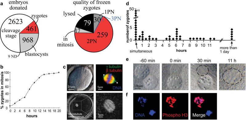
Figure 1. Donation of human zygotes for stem cell research.
a, embryos donated for stem cell research. PN= pronucleus, ND= not determined b, Mitotic entry time of human zygotes (n=107) in hours after thaw. Nuclear envelope breakdown of at least one of the two pronuclei was defined as the time point of mitotic entry. Some zygotes were mitotic at the time of thaw. c, mitotic spindle formation. Human zygote 30 minutes after nuclear envelope breakdown including brightfield image, microtubule immunohistochemistry (arrowheads point to the centrosome at both poles of the spindle) and spindle birefringence. d-f, asynchrony in nuclear envelope breakdown. d, Hours between the breakdown of the first pronuclear envelope and the second. e, Zygote with asynchronous pronuclear envelope breakdown. Numbers indicate the time from the breakdown of the first nuclear envelope. The location of mitotic chromatin is circled. f, 1PN zygote stained for phosphorylation of Ser 27 on Histone H3, a marker of mitosis.

