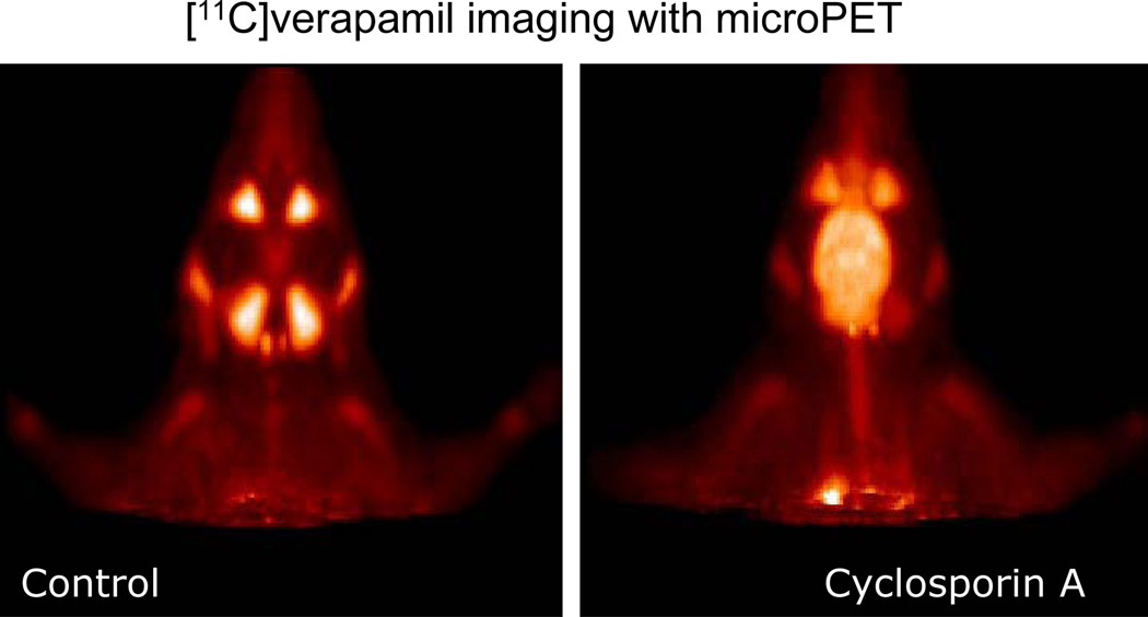
Figure 4. Micro PET images of the head biodistribution of calcium channel blocker.
Micro PET images of the head of a Wistar rat, showing the biodistribution of the calcium channel blocker, [11C] verapamil injected systemically, either alone (Control) or after pre-treatment of the animal with the Pgp inhibitor Cyclosporin A. [11C] verapamil, a substrate for the blood-brain barrier efflux transporter P-glycoprotein, gains access to the brain only after Pgp inhibition by Cyclosporin A. Images are courtesy of Dr. P. Elsinga, University Medical Center Groningen, The Netherlands.

