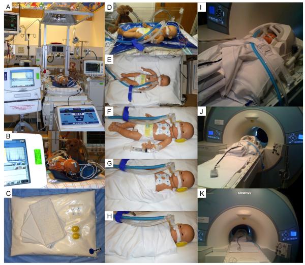
FIGURE 1.
Procedure for performing brain MRI in neonates with hypoxic-ischemic encephalopathy treated with induced hypothermia. A-B: During hypothermia treatment neonates are maintained on the hypothermia blanket and hypothermia system in the incubator with the canopy up and the heat source off. During the whole treatment, neonates are monitored by amplitude-integrated electroencephalogram. C. Materials: a MRI-compatible pillow containing Styrofoam and 1-2 thin blankets, stored for a few hours prior to the exam in a 4°C fridge, as well as earmuffs and complete MRI-compatible cardiovascular monitoring. D-H: Neonates are wrapped with 1-2 thin blankets and placed on a MRI-compatible pillow containing Styrofoam. Ears are covered with earmuffs. The MRI-incompatible esophageal probe is removed. Complete MRI-compatible cardiovascular monitoring is placed. I-K: Once the neonate is in place in the MRI scanner, the air in the MRI-compatible pillow is suctioned, and imaging process starts.

