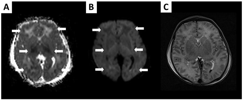
FIGURE 3.
Brain MRI performed on day of life 2 in a newborn demonstrating total cortical injury pattern while he is still receiving therapeutic hypothermia; (A) ADC map, (B) DWI images, and (C) T2-weighted imaging. Clear diffusion abnormalities are present in the cortex, white matter and basal ganglia of this newborn as seen on ADC map and DWI images (arrows), while findings are not so evident on concomitant T2-weighted imaging.

