Figure 2.
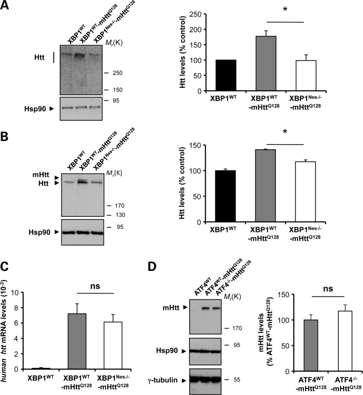
XBP1 deficiency in the nervous system decreases mHtt levels in the YAC128 HD model. (A) Levels of Huntingtin (Htt) were measured in the striatum of 6-month-old mice by western blot analysis using the anti-Htt clone mEM48 antibody. XBP1WT animals were analyzed as control. Hsp90 was monitored as loading control. Htt levels were quantified in striatum extracts from XBP1WT (n= 3), XBP1WT-mHttQ128 (n= 8) and XBP1Nes−/−-mHttQ128 (n= 13) mice (left panel). Mean and SEM are presented. (B) In parallel, Htt relative levels were determined using anti-Htt clone 1HU-4C8 antibody. Htt levels were quantified in the brain striatum of XBP1WT (n= 3), XBP1WT-mHttQ128 (n= 3) and XBP1Nes−/−-mHttQ128 (n= 3) mice (left panel). Mean and SEM are presented. *P< 0.05, calculated with Student's t-test. (C) The mRNA level of the human huntingtin gene was analyzed by real-time PCR in total cDNA obtained from the brain striatum of XBP1Nes−/−-mHttQ128 or littermate control mice. All samples were normalized to β-actin levels. Average and SEM of the analysis of three animals per group are shown. (D) mHtt levels were analyzed using anti-polyQ clone 3B5H10 antibody in the striatum of 12-month-old mice (upper panel). Right panel: mHtt levels were quantified in ATF4WT (n= 3), ATF4WT-mHttQ128 (n= 4) and ATF4−/−-mHttQ128 (n= 4) (bottom panel). In addition to Hsp90, γ-tubulin levels were analyzed as loading control. In all experiments, littermates were employed for comparison.

