Figure 6.
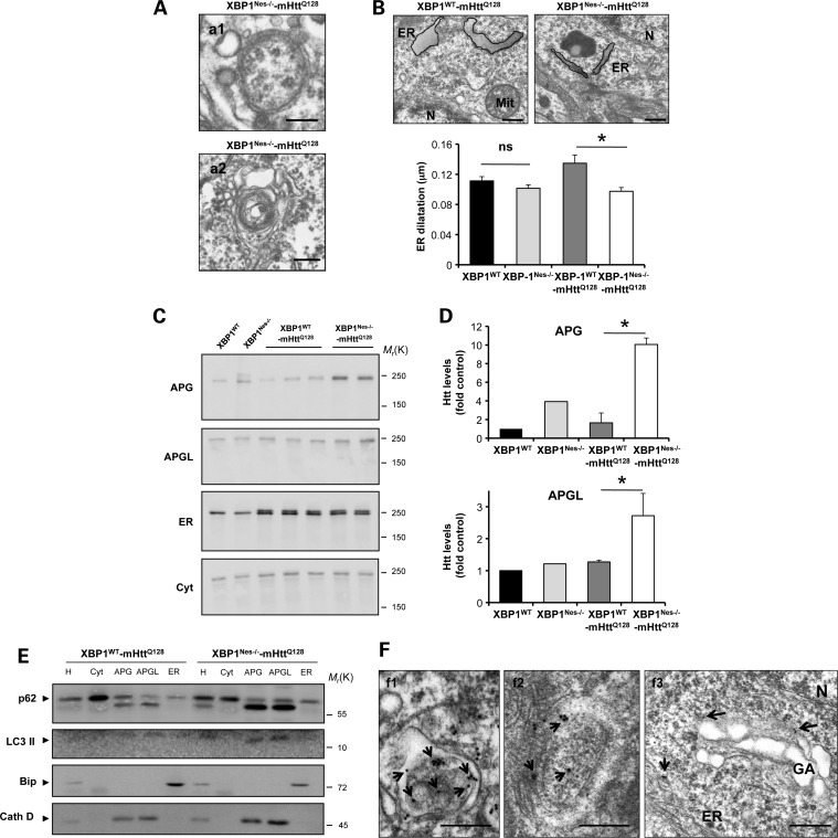
Loss of XBP1 targets mHtt to the autophagy pathway. (A) The accumulation of APG-like structures was visualized by EM in neurons of the striatum of XBP1Nes−/−-mHttQ128 mice at 6 months of age. Scale bars, 230 nm (a1) and 600 nm (a2). Images represent the analysis of three independent animals. Neurons were identified by their morphology at low magnification. (B) ER structure was visualized by EM in neurons of the striatum of mice at 6 months of age. Scale bar, 300 nm. Left panel: ER dilatation was quantified from XBP1WT (n= 3), XBP1Nes−/− (n= 3), XBP1WT-mHttQ128 (n= 3) and XBP1Nes−/−-mHttQ128 (n= 3) mice as described in Materials and Methods. Mean and SEM are presented. As an example, the perimeter of the ER was marked with a black line with a gray area. *P<0.05 calculated with Student's t-test. (C) Brain extracts from indicated animals were subjected to subcellular fractionation to purify fractions enriched in different organelles including autophagosomes (APG), autophagolysosomes (APGL), ER and cytosol (cyt). (D) Quantification of Htt levels in the autophagic compartments relative to XBP1WT is shown (XBP1WT-mHttQ128, n= 3; XBP1Nes−/−-mHttQ128, n= 3) using the anti-Htt clone 1HU-4C8 antibody. Mean and SEM are presented. (E) Biochemical characterization of the fractions isolated in (C). Immunoblot for the indicated marker proteins to verify the purity of the fractions was performed for the homogenate (H), cytosol (cyt), autophagosomes (APG), autophagolysosomes (APGL) and endoplasmic reticulum (ER) isolated from cortex and midbrain. (F) Immunogold staining and EM analysis of the brain striatum, using an anti-expanded polyQ antibody (clone 3B5H10) was performed in tissue derived from XBP1Nes−/−-mHttQ128 mice. (f1) Co-localization with ER membrane. Scale bar, 500 nm. (f2) A double-membrane APG-containing positive immunogold staining in its lumen. Scale bar, 300 nm. (f3) An example of co-localization of immunogold staining and Golgi apparatus membrane is shown. Scale bar, 500 nm. Data represent the analysis of three independent animals. Arrows indicate immunogold staining. N, nucleus; GA, Golgi apparatus; ER, endoplasmic reticulum.

