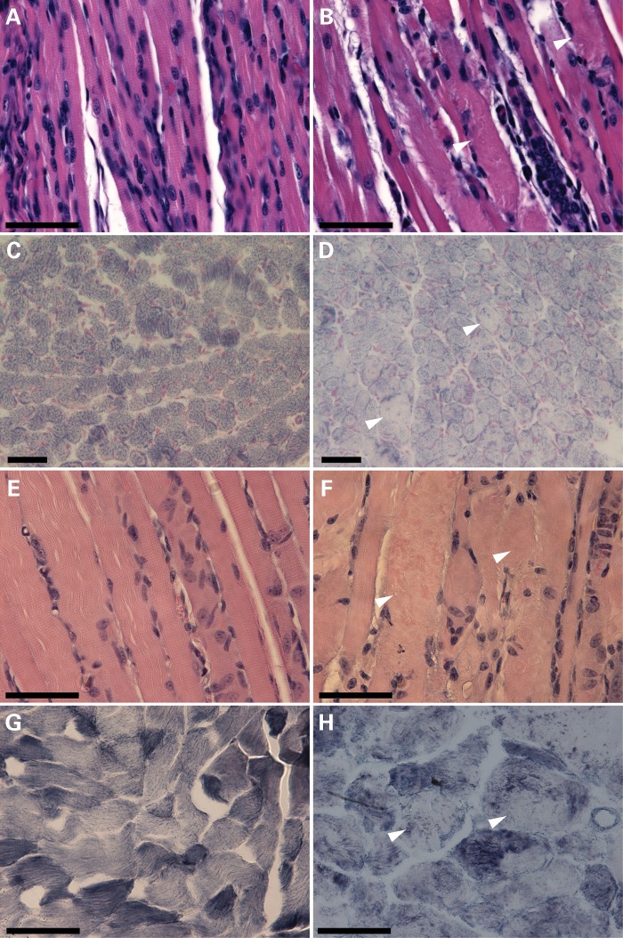
Figure 3.
Skeletal muscle histopathology of cofilin-2-deficient mice (Cfl2−/− and Cofi/Cofi:Ckmm+). H&E staining (A, B, E and F) of quadriceps cross-sections from WT (A) and Cfl2−/− (B) at P7, and Cofi/+:Ckmm+ (E) and Cofi/Cofi:Ckmm+ (F) mice at P21 was performed. Sections from Cfl2−/− and Cofi/Cofi:Ckmm+ muscles revealed ballooning sarcomeric degeneration (arrowheads). NADH staining (C, D, G and H) was performed on quadriceps cross-sections from WT (C), Cfl2−/− (D), Cofi/+:Ckmm+ (G) and Cofi/Cofi:Ckmm+ (H) mice. Sections from Cfl2−/− and Cofi/Cofi:Ckmm+ mice revealed pale areas within the myofibers as shown by arrowheads (scale bar = 50 μm).

