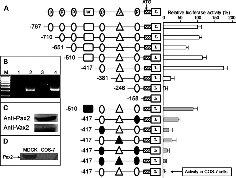
Figure 2.
pax2.1 responsive elements in the fadd promoter. (A) Diagram at top depicts symbols used for transcription factor-binding sites. p, pax2.1 site; v, vax2 site; rxr, rxr site. Hatched box, exon 1 of fadd. L, luciferase gene. Reporter constructs transfected into Madin-Derby canine kidney (MDCK) cells. In lower part of (A), the filled symbols represent mutation of the site so it is no longer functional. The activity of the last construct relates to the −417 bp fragment transfected into COS-7 cells. The relative luciferase activity for the longest construct (−767 bp) was set at 100%. Activity (± SEM, n= 6) of all other constructs measured against this. (B) Confirmation that MDCK cells express Rxr. Lane 1, no RT control for Rxr-α; lane 2, Rxr-α (495 bp); lane 3, no RT control Rxr-β; lane 4, Rxr-β (520 bp). M, 100 bp marker. (C) Western blot of MDCK cells with Pax2 or Vax2 antibodies. (D) Western blot showing the Pax2 protein is absent in COS-7 cells.

