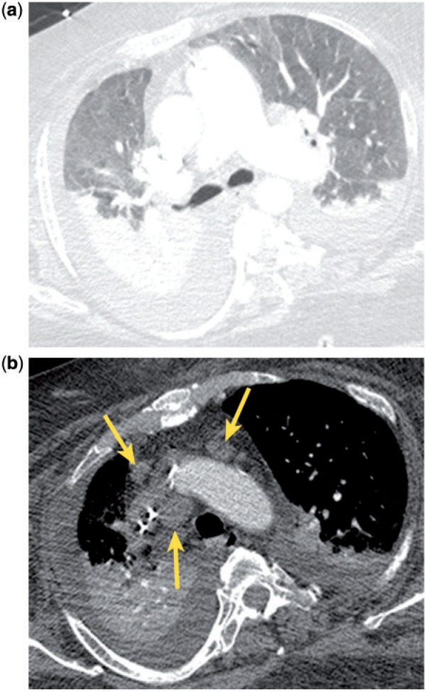
Figure 2.

Incidental mediastinal lymph node enlargement in a patient with CHF. CT shows ground glass pulmonary opacities, parenchymal consolidation and smooth septal thickening (a), as well as bilateral pleural effusions, consistent with CHF. There are transvenous pacemaker leads in the superior vena cava and enlarged mediastinal lymph nodes (b, arrows).
