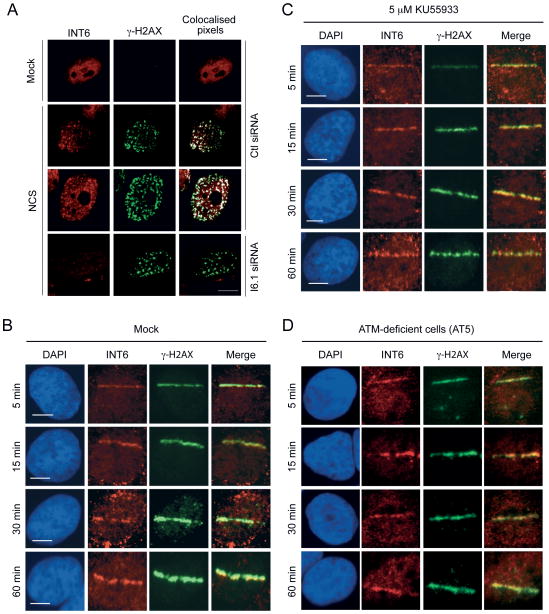
Figure 4.
INT6 is recruited to DSBs induced by NCS treatment or micro-irradiation. (A) HeLa cells transfected with control or INT6-targeting siRNAs were treated, or not, with NCS (200 ng/ml, 15 min), and immunostained 3 h later with antibodies against INT6 and γ-H2AX. Representative confocal images are shown. Co-localized pixels were determined using the Co-localization Highlighter plug-in for ImageJ software. They appear as white dots on right panels. Scale bar, 10 μM. (B and C) U2OS cells were untreated (B) or pre-treated with 5 μM KU55933 (C), micro-irradiated, fixed at the indicated time points, immunostained with antibodies to INT6 and γ-H2AX, and counterstained with DAPI. The merged red and green channels show co-localization in yellow. Bar, 10 μM. (D) Recruitment of INT6 was assessed in ATM-deficient cells (AT5) processed as above.

