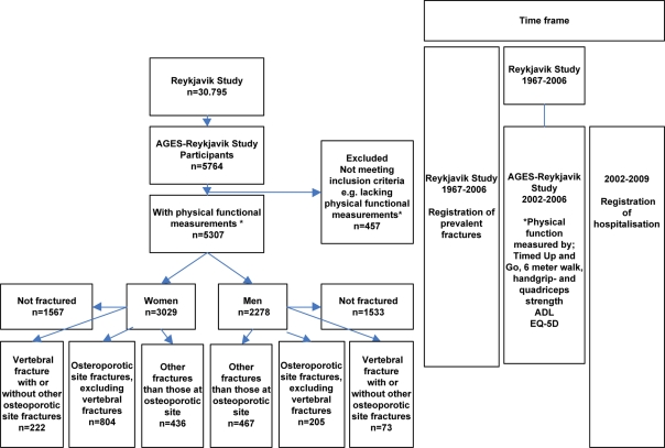
Figure 1.
Flow chart of timeline and participants categorised by sex and previous fractures. Fractures sustained before the entry into the AGES-Reykjavik Study were recorded, verified and confirmed from medical and radiological records as described [16]. The following were defined as osteoporotic sites: vertebral (S12.1, S12.2, S22.0, S22.1, S32.0, T08), pelvic (S32.1, S32.3, S32.4, S32.5), proximal humerus (S42.2, S42.4), distal forearm (S52.5, S52.6), hip (S72.0, S72.1, S72.2), proximal tibia (S82.1).

