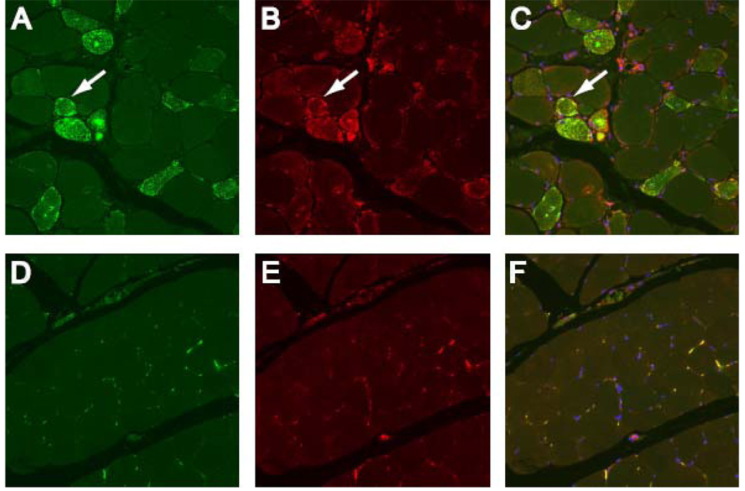
Figure 4.
HMGCR expression is up-regulated in regenerating myofibers expressing NCAM. Muscle biopsies from anti-HMGCR positive (A, B, and C) and control subjects (D, E, and F) were co-stained with anti-NCAM (A and D; green), anti-HMGCR antibodies (B and E; red) and DAPI (blue) to stain nuclei. Overlay (C and F) demonstrates HMGCR and NCAM are frequently co-expressed at high levels in the same myofibers in anti-HMGCR positive biopsies (white arrows) but not control muscle tissue. To ensure comparability, images A–C and D–F were obtained using identical exposure settings for each channel. These results are representative of staining seen in 6 anti-HMGCR positive and 3 normal muscle biopsies. (20X objective).

