Figure 1.
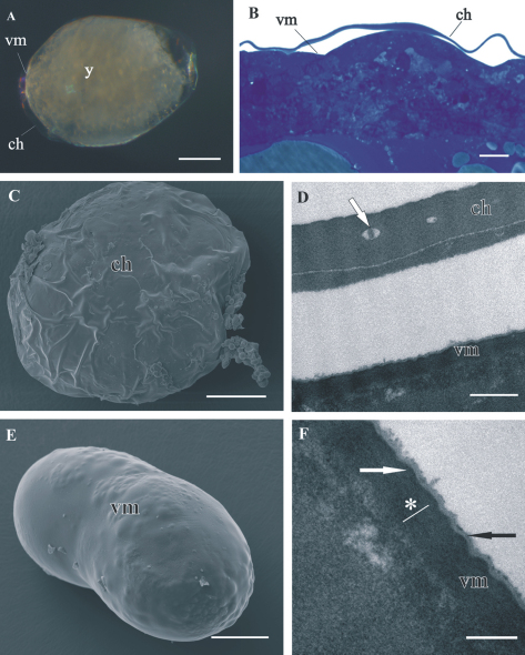
Structure of distal chorion (ch) and proximal vitelline membrane (vm), covering Porcellio scaber A, B, D, F and Porcellio dilatatus C, E early-stage embryo. A The early-stage embryo with large amount of yolk (y) and no visible limb buds. B Semithin section of the embryo peripheral region. Chorion is separated from the embryo surface. The vitelline membrane is closely apposed to the embryo surface. C SEM micrograph of the early-stage embryo. The outer egg envelope, chorion, is visible. D TEM micrograph of one-layered chorion, including electron lucent “lacunae” (white arrow). There is a layer of artificially spilt yolk underneath the chorion. E SEM micrograph of the early-stage embryo. Chorion is artificially removed and the inner egg envelope, vitelline membrane, is exposed. F TEM micrograph of vitelline membrane, composed of three layers: main proximal homogenous layer (*), thin middle electron dense layer (white arrow) and superficial corrugated lucent layer (black arrow). Bars: A, C, E 200 µm; B 10 µm; D 0.5 µm; F 200 nm.

