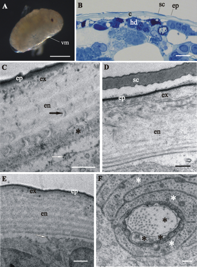
Figure 4.
Cuticle structure and renewal in Porcellio scaber prehatching late-stage embryo. A Swelled embryo inside the vitelline membrane (vm), prior to hatching. B Semithin section of the embryo peripheral region. The vitelline membrane is artificially removed. Clearly discernible exoskeletal cuticle (c), detached from the underlying hypodermis (hd). C, D, E TEM micrographs of exoskeletal cuticle in different regions of the same specimen, composed of three principal layers: the outermost thin electron dense epicuticle (ep), the middle exocuticle (ex) and the innermost endocuticle with several sublayers (en). The micrographs show features of cuticle renewal: cuticle detachment from the hypodermis, partial disintegration of proximal portion of endocuticle (*) and irregularly arranged electron dense particles on outer apical plasma membrane surface (white arrows). Pore canals (black arrow) in the endocuticle consist of electron lucent central part and electron dense margins C. Cuticular scales (sc) are fully elaborated and the exocuticle has the characteristic pattern of chitin-protein fibers arrangement D. Exocuticle is hardly discernible E. F TEM micrograph of completely structured sensillum transverse section in the hypodermis. Dendritic outer segments (*) and enveloping cells (white *). Bars: A 500 µm; B 10 µm; C, E 1 µm; D 0.5 µm; F 200 nm.

