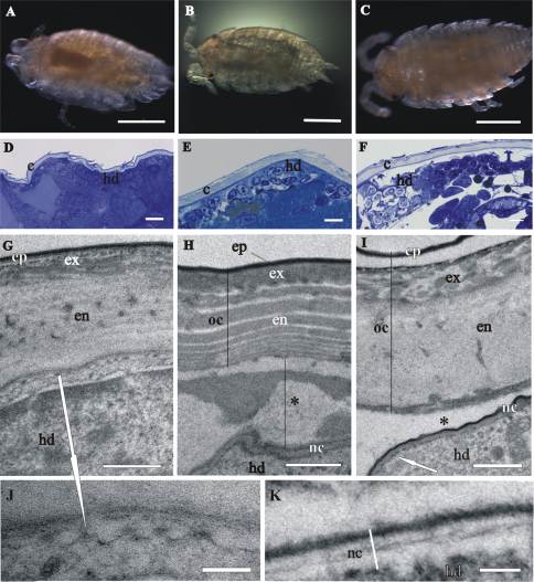
Figure 5.
Cuticle structure and renewal in Porcellio scaber marsupial mancas. A The early-stage marsupial manca, immediately after hatching B The mid-stage marsupial manca C The late-stage marsupial manca, just prior to release from the marsupium D–F Semithin sections of the manca peripheral region in the early-stage marsupial manca D in the mid-stage marsupial manca E and in the late-stage marsupial manca F. Cuticle (c), overlying the hypodermis (hd), becomes progressively more similar to adult cuticle. G–K TEM micrographs of exoskeletal cuticle in the early-stage marsupial manca G, J in the mid-stage marsupial manca H and in the late-stage marsupial manca I, K Three main layers are distinguished: epicuticle (ep), exocuticle (ex) and endocuticle (en). The micrographs show morphological characteristics of cuticle renewal: detachment of the old cuticle (oc) from the hypodermis, ecdysal space (*) between the detached cuticle and the newly forming cuticle (nc) and partial degradation of the old cuticle H, I protrusions with electron dense tips (white arrows) on apical surfaces of hypodermal cells G, I, J. The new cuticle consists of two layers, external electron dense epicuticle and internal electron lucent procuticle H, I, K. Helicoidal chitin-protein fibers arrangement is discernible in some regions of late-stage marsupial manca K. Bars: A–C 500 µm; D–F 10 µm; G–I 1 µm; J, K 200 nm.

