
Figure 3.
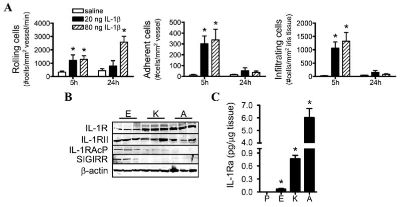
Investigation of the eye’s responsiveness to interleukin (IL)-1β. (A) Wild-type (WT) mice were administered an intraocular injection of the indicated concentrations of recombinant IL-1β or saline, and the cellular inflammatory responses in the iris were assessed by intravital microscopy. The numbers of rolling, adhering and infiltrating cells were quantified (*p<0.05 for comparison between saline and IL-1β injections; n=8–10 mice/treatment/time). (B) Immunoblotting of tissue homogenates from naïve, WT mice comparing expression levels of IL-1R and the indicated negative regulators of IL-1 across the three tissues. Shown are immunoblots of three mice; a total of nine mice were examined. (C) The concentrations of IL-1 receptor antagonist (IL-1Ra) in naïve mice were measured by ELISA. *p<0.05 for comparison with plasma concentrations. P, plasma; E, eye; K, knee; A, ankle. IL-1RAcP, IL-1 receptor accessory protein; SIGIRR, single Ig IL-1R-related molecule.