
Figure 4.
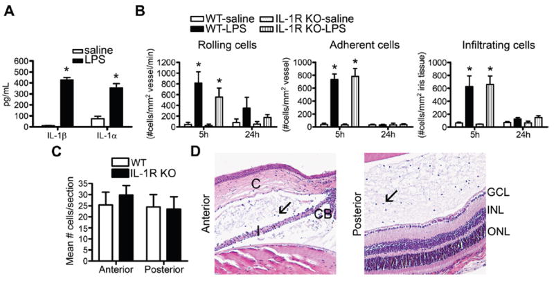
Interleukin 1 receptor (IL-1R) deficiency does not alter the severity of endotoxin-induced uveitis (EIU). (A) IL-1β and IL-1α concentrations in eye homogenates of wild-type (WT) mice were analysed by ELISA to compare responses to intraocular injection of 250 ng LPS vs saline 5 h after injection. WT and IL-1R knockout (KO) mice were administered an intraocular injection of 250 ng LPS or saline, and the severity of uveitis was assessed by intravital microscopy as a function of time (B) or histologically (C, D) at 24 h after LPS injection. (D) depicts images of the anterior and posterior eye segments of IL-1R KO mice. Arrows indicate leucocytes in the aqueous humour of the anterior chamber or the vitreous body of the posterior chamber. CB, ciliary body; C, cornea; I, iris; ONL, outer nuclear layer; INL, inner nuclear layer; GCL, ganglion cell layer. *p<0.05 for comparison between LPS and saline (n=14–17 mice/treatment/genotype). No significant difference among genotypes was observed. Results are combined data from two independent experiments.