
Figure 6.
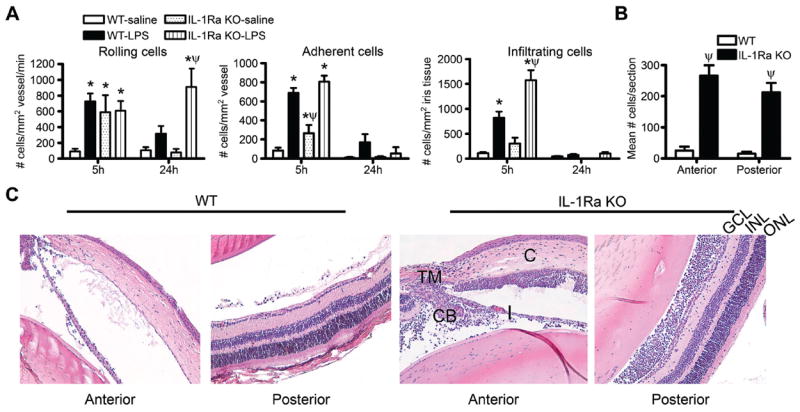
Interleukin 1 receptor antagonist (IL-1Ra) deficiency renders mice more susceptible to locally administered lipopolysaccharide (LPS). Wild-type (WT) or IL-1Ra knockout (KO) mice were administered an intraocular injection of 250 ng LPS or saline, and the cellular inflammatory response in the iris was assessed by intravital microscopy. The numbers of rolling versus adherent versus infiltrating cells were quantified as a function of time (A; *p<0.05 for comparison between LPS responses vs saline within a genotype; ψp<0.05 for comparison between LPS response in WT vs IL-1Ra KO). The extent to which IL-1Ra regulates uveitis was assessed histologically at 24 h after injection, and the numbers of infiltrating leucocytes in the anterior or posterior eye segments after LPS injection was quantifi ed (B; ψp<0.05 for comparison between LPS response in WT vs IL-1Ra KO; both WT and IL-1Ra KO mice showed significant responses over saline-injection controls). (C) shows representative histological images of WT versus IL-1Ra KO eyes at 24 h after LPS injection. C, cornea; I, iris; TM, trabecular meshwork; ONL, outer nuclear layer; INL, inner nuclear layer; GCL, ganglion cell layer. n=10–16 mice/genotype/treatment/time. Results are combined data from two independently performed experiments.