
Figure 7.
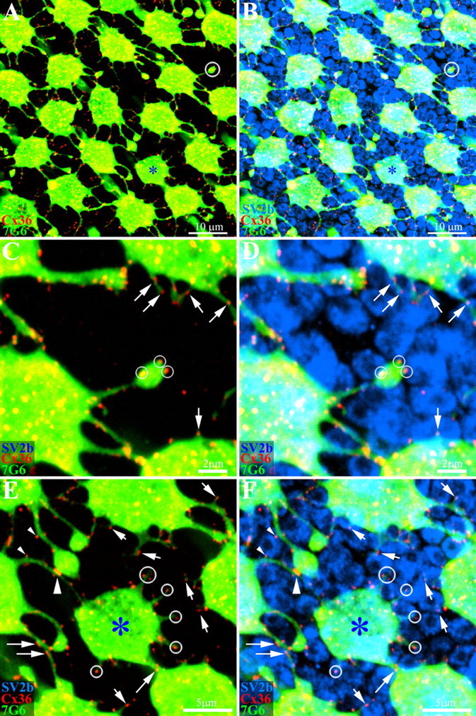
Primate cones make Cx36 gap junctions with rod spherules. Whole-mount macaque retina was stained with antibodies against cone arrestin (7G6, green), Cx36 (red) and SV2b (blue), to stain rod spherules. A, A mosaic of cone pedicles in peripheral retina. A blue cone pedicle is marked with an asterisk. As before, there are many Cx36 plaques at telodendrial contacts. In addition, a few telodendria terminate in isolation (circle, top right). B, The same field shows that the space between cone pedicles is mostly filled by rod spherules stained for SV2b. C, High-magnification image of the isolated telodendron from A shows clear Cx36 plaques, even in the absence of adjoining cones pedicles (small circles). In addition, many other small telodendria, also bearing Cx36 plaques, project from a cone pedicle base (arrows). D, The isolated cone telodendron is surrounded by rod spherules and the Cx36 plaques occur at contact points with rod spherules (small circles). The Cx36 plaques on short telodendria also contact rod spherules (arrows). E, The blue cone pedicle also has many fine telodendria, too short to reach adjacent cones. One that approaches a nearby cone has Cx36 plaques aligned with rod spherules (diagonal arrow). In addition, Cx36 plaques may be observed along a telodendrial shaft (long arrows). F, Cx36 plaques on the blue cone telodendria are aligned with the surrounding rod spherules (circles). Some rod spherules have Cx36 contacts with the blue cone pedicle and an adjacent cone pedicle. Two small Cx36 plaques from different telodendria contact opposite sides of a single rod spherule (small arrowheads). Further down, there is a much larger cone-to-cone gap junction (large arrowhead). In summary, many fine telodendria form all types of cones make small Cx36 gap junctions with surrounding rod spherules (small arrows). A–D, 4 × 0.4 μm optical sections; E, F, 3 × 0.4 μm optical sections.