Figure 4.

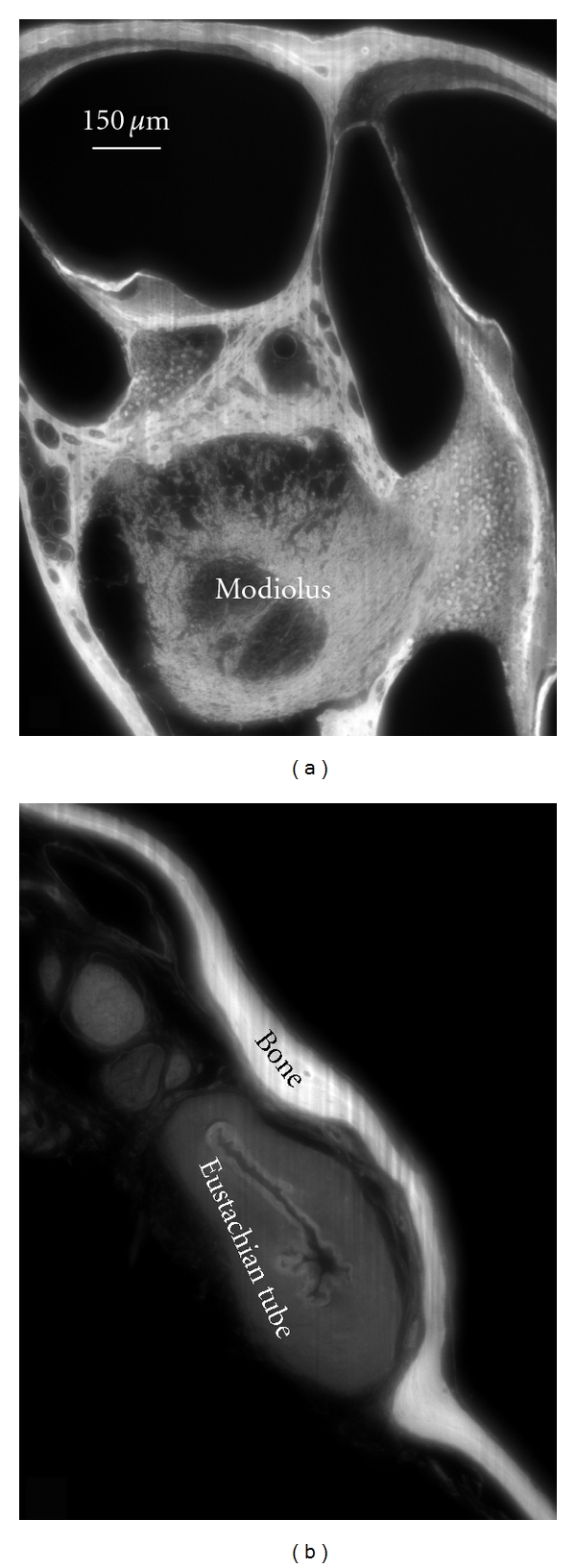
An OPFOS cross-section of 1600 × 1200 pixels through (a) the scalae and modiolus of a gerbil inner ear cochlea and (b) a closed Eustachian tube in the middle ear. Rhodamine staining was combined with 532 nm laser light sheet sectioning.

An OPFOS cross-section of 1600 × 1200 pixels through (a) the scalae and modiolus of a gerbil inner ear cochlea and (b) a closed Eustachian tube in the middle ear. Rhodamine staining was combined with 532 nm laser light sheet sectioning.