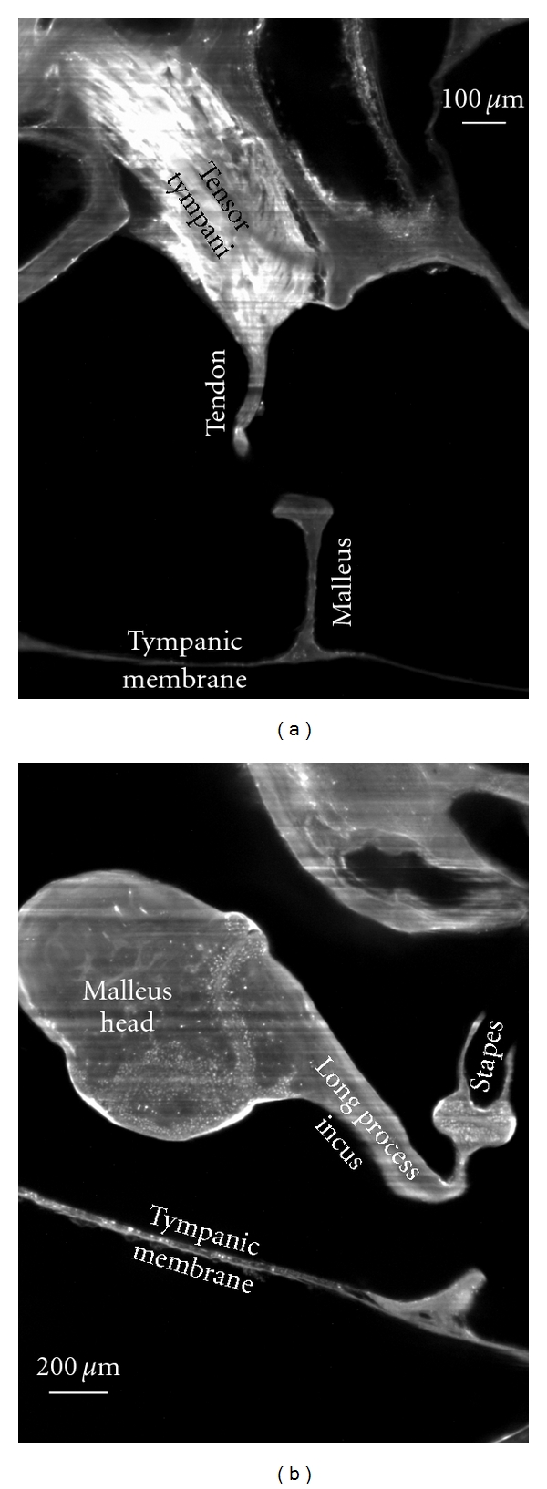
Figure 5.

2D virtual cross-sections (1600 × 1200 pixels) from OPFOS microscopy on the gerbil middle ear. (a) Tensor tympani muscle and tendon reaching down towards the malleus hearing bone. (b) Incudomalleolar and incudostapedial articulation between incus and malleus hearing bone. Rhodamine staining was combined with 532 nm laser light sheet sectioning. Pixel size 1.5 × 1.5 μm.
