
Figure 1.
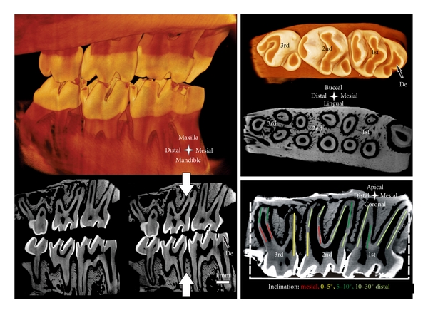
Left top: Simulated occlusion of a left rat maxilla with the corresponding mandible imaged with Micro-XCT. Left bottom: sagittal sections simulate open (left) and closed (right) bite. Top right: occlusal surface and transversal section of a right maxilla. Bottom right: sagittal section; note the predominant distal inclination of the roots. Inclination α is measured for the most mesial root as demonstrated. The colored segments on the roots represent the inclination angle of the roots. De = exposed dentin.