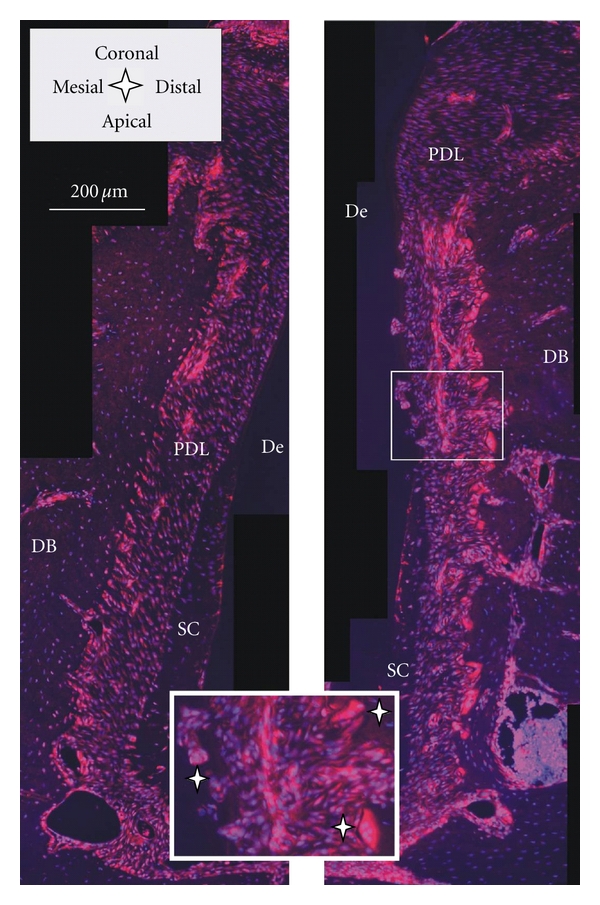
Figure 8.

Histological sections immunostained with RANKL; the red RANKL stain is dominant in the PDL close to the bone surface of the distal root-bone complex (right image) compared to the mesial complex (left image); insert: note odontoclast on root and osteoclasts on bone (white stars). De = dentin, DB = interdental bone, PDL = periodontal ligament, and SC = secondary cementum.
