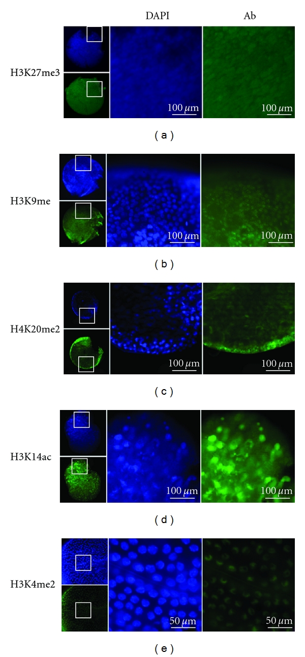
Figure 2.

Whole mount immunocytochemistry of Daphnia magna embryos stained with antibodies specific to modified histone H3 and H4. The small images show the embryo from which the magnified images to the right are shown. Blue staining is with DAPI, which detects all DNA. Green staining (Ab) is for the specified histone modification. (a) H3K27me3. (b) H3K9me. (c) H4K20me2. (d) H3K14ac. (e) H3K4me2. The embryos shown in (a–d) are blastula stages, (e) is a magnified view of a gastrula embryo. The low magnification views show the torn extraembryonic membranes required to allow antibody penetration to the cells.
