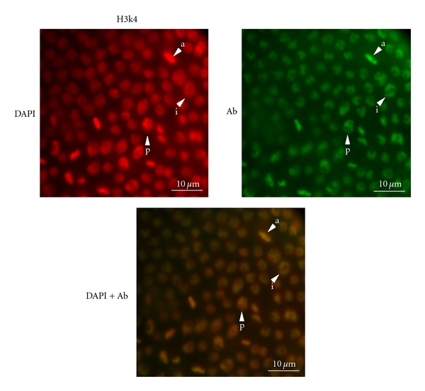
Figure 3.

Localized, cell-cycle-dependent H3K4me2 staining in Daphnia magna gastrula nuclei. In interphase cells (i), the DAPI staining (red, left) is largely uniform whereas the H3K4me2 staining (green, right) is concentrated at the nuclear periphery producing a yellow-green circle with a red center in the merged image (lower panel). In cells undergoing prophase (p) and anaphase (a) the DAPI and H3K4me2 staining is coincident. Multiple cells in these stages are shown.
