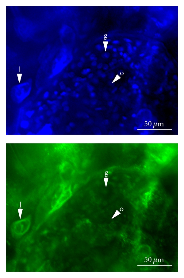
Figure 4.

Whole mount immunocytochemistry of Daphnia magna ovaries stained with antibodies specific to H3K4me2. The top image shows DAPI staining, which detects all nuclei including the large lipid cell (l), the nuclei of the germarium (g), present in a rosette arrangement, and the nuclei of the developing oocytes and nurse cells (o). The lower image shows the same tissue stained for H3K4me2. The nuclei of the lipid cell and the cells of the germarium are detected. The nuclei of the oocytes and nurse cells are not strongly labelled with this antibody.
