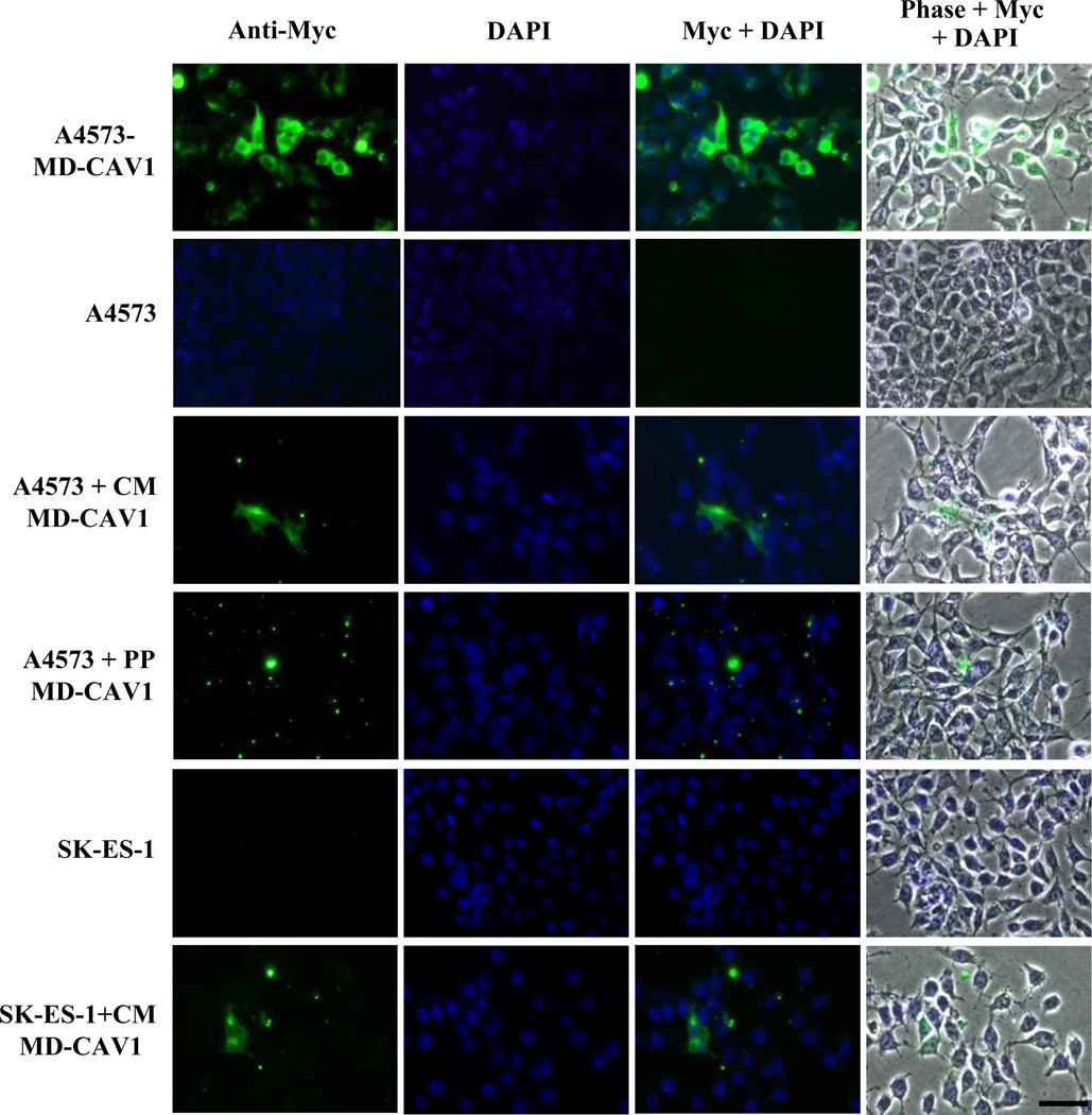
Figure 4. Uptake of human CAV1 by EWS cells.
Immunofluorescent detection of CAV1 taken up by A4573 or SK-ES-1 cells either from conditioned medium (CM MD-CAV1) or from culture medium supplemented with the purified protein (PP MD-CAV1). MD-CAV1 was visualized using an Alexa Fluor 488 (green)-conjugated secondary antibody following immunodetection of the Myc tag, while the nuclei were visualized using DAPI (blue). The panels show individual channels detecting the protein (Anti-Myc) and nuclei (DAPI) along with fluorescent merged channels (Myc + DAPI) and fluorescent channels merged with phase contrast image (Phase + Myc + DAPI). The size bars corresponds to 50µm.

