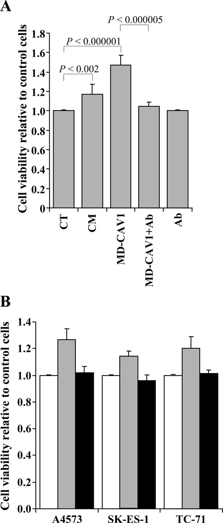
Figure 5. Exogenously added CAV1 stimulates cell proliferation in EWS cells.
(A) Proliferation of A4573 cells was measured using a luminescence-based technique in untreated cells (CT), and in cells incubated with conditioned medium containing CAV1 (CM); with exogenously added purified MD-CAV1; with exogenously added purified MD-CAV1 along with anti-CAV1 antibody (MD-CAV1 + Ab); and with the anti-CAV1 antibody alone (Ab). (B) Comparison of proliferation of EWS cell lines in the presence of either exogenously added MD-CAV1 (gray bar) or exogenously added MD-CAV1 plus the anti-CAV1 polyclonal antibody (black bar), with the control cells (white bar). Cell viability values in both panels are plotted relative to those in untreated control cultures, which were arbitrarily set as 1.00.

