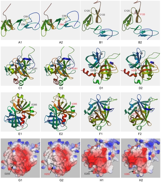
Figure 2. Structure models of the wild-type and mutant PC proteins.
The amino acid number was designated according to the previous nomenclature described in the Human Gene Mutation Database. That is, the first 42 amino acids (signal peptide) is subtracted. The wild-type amino acids are shown in yellow and the mutants are shown in red. The D255H and the E285V mutations are also displayed in a solid surface model (G, H) in which the electrostatic potential is clearly indicated.

