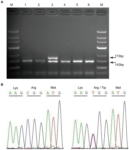
Figure 3. PROC c.565C>T variant detection by PCR-RFLP and direct sequencing.
(A) Electrophoretic patterns following Hin6 I digestion. PCR products were 173 bp. Only amplicons with the wild-type sequences were digested, yielding two bands of 143 bp and 30 bp. The digestion products were separated by 2% agarose gel electrophoresis. M, DNA marker with 50-bp ladder. Lanes 1, 2, and 4–6, normal individuals. Lane 3, heterozygous individual for the variant. (B) Chromatograms obtained by sequencing. Left, wild-type; Right, heterozygote.

