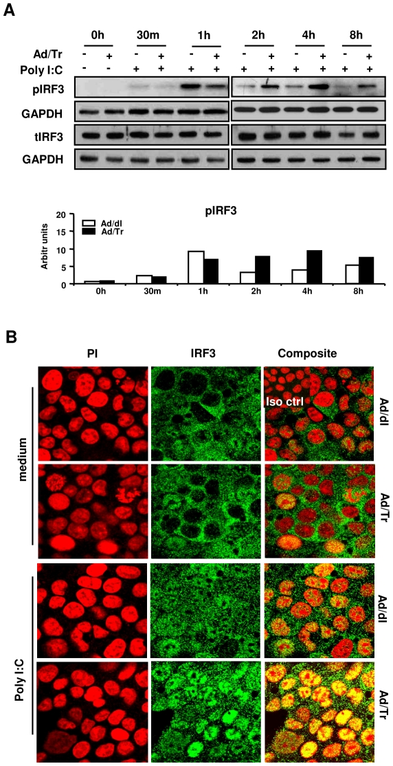
Figure 4. Enhanced polyI∶C-driven antiviral protection in Ad/Tr-cells is associated with hyperactivation of IRF3.
(A) Western blots and their analyses of phosphorylated IRF3, total IRF3, and GAPDH proteins were performed from whole-cell extracts of HEC-Ad cells that were either left untreated or treated with 25 µg/ml polyI∶C during indicated time points. (B) Immunofluorescence analysis of IRF3 nuclear translocation following either medium alone or polyI∶C 25 µg/ml treatment for 4 h. Representative staining is shown for IRF3 (green), nuclear stain (PI) (red), and composite (yellow) at magnification 2520×. The data are representative of three independent experiments with similar results.

