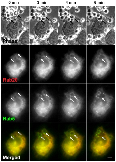
Figure 2. The association of Rab20 with phagosomal membranes persists following the loss of Rab5.
Live RAW264 cells co-expressing CFP-Rab20 (red) and GFP- Rab5 (green) were incubated with IgG-Es and monitored by phase-contrast and fluorescence microscopy. The elapsed time is indicated at the top. The binding of IgG-Es to the cell surface was set as time 0. It was found that Rab20 and Rab5 were transiently colocalized on formed phagosomes and that Rab20 remained associated with phagosomal membrane from which Rab5 was dissociated (arrows). Representative images from three independent experiments are shown. Scale bar: 5 µm. The corresponding video is Movie S2.

