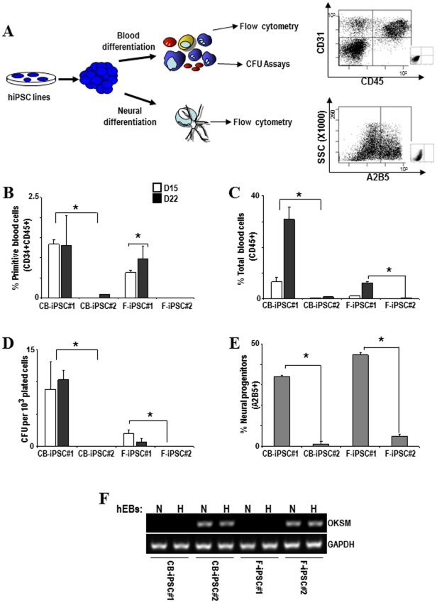
Figure 3. Residual expression of ectopic reprogramming factors prevents differentiation of hiPSC lines.
(A) Schematic of the hematopoietic and neural differentiation protocol from hiPSC and endpoint analyses. Right panel: representative flow cytometry dot plots displaying how primitive hematopoietic cells (CD45+CD34+), mature hematopoietic cells (CD45+) and cells committed towards early neuroectoderm (A2B5+) are identified and analyzed. (B–E) Lines CB-hiPSC #2 and F-hiPSC #2 display a significant impairment in their differentiation capacity into primitive blood cells (CD45+CD34+) (B), total blood cells (CD45+CD34−) (C), colony-forming unit potential (D) and early neural progenitors (E). (F) The expression of the reprogramming factors remain upon hematopoietic (H) and neural (N) differentiation, consistent with that observed in undifferentiated hiPSC lines.

