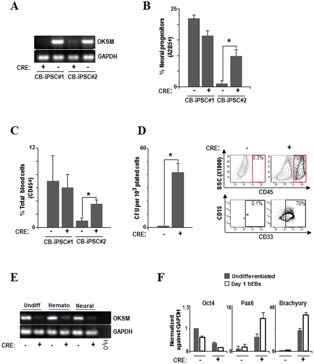
Figure 4. Transgene-free hiPSC clones display improved differentiation capacity.
(A) PCR of genomic DNA (gDNA) showing successful adenovirus-based Cre-excision of the reprogramming cassette in CB-hiPSC. CB-hiPSC #2 after Cre-excision significantly improved its differentiation capacity into early neural progenitors (B) total blood cells (C) and colony-forming unit potential (D). Flow cytometry from CB-hiPSC #2 CFUs before and after Cre excision clearly shows that CD45+ and CD45+CD33+ myeloid cells appear in CFUs from CB-hiPSC #2 after Cre excision. (E) RT-PCR confirming that in CB-hiPSC #2 after Cre-excision the expression of ectopic reprogramming factors is greatly reduced in undifferentiated hiPSC and after 15 days of hematopoietic and neural differentiation. (F) Q-RT-PCR showing an increased expression of the early differentiation markers, PAX6 (neural) and Brachyury (mesendoderm), in CB-iPSC #2 after Cre-mediated OKSM excision. Q-RT-PCR expression is normalized against GAPDH.

