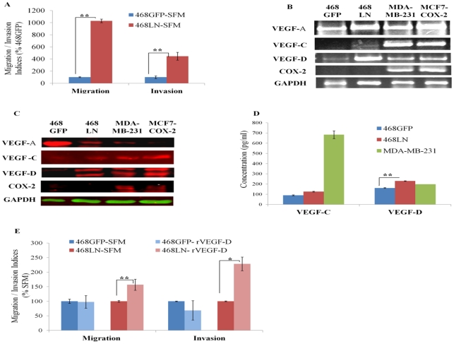
Figure 1. Differential migratory, invasive, and VEGF-D producing capacity of 486LN cells, as compared to 468GFP cells:
(A) Compared to 468GFP cells, 468LN cells were significantly more migratory and invasive. (B, C) 468LN cells expressed significantly higher level of VEGF-D mRNA measured with semi-quantitative RT-PCR (B) and total protein measured with western blot (C), compared to 468GFP cells. (MDA-MB-231 and MCF7-COX-2 cells served as positive controls for COX-2 and VEGF-C/D). (D) 468LN cells secreted significantly higher levels of VEGF-D but not VEGF-C, in comparison to 468GFP cells as measured by ELISA in cell supernatants; MDA-MB-231 cells served as positive controls for both VEGF-C and VEGF-D. (E) Exogenous rVEGF-D (2.5 ng/ml) increased both migration and invasion of 468LN, but not 468GFP cells. Migration/ invasion indices were normalized relative to 468GFP in SFM. Migration and invasion of 468GFP-SFM in Fig E was performed separately from the data in Fig A. All bars represent mean (n = 4) +/− S.E, *, P< 0.05; **, P< 0.01.

