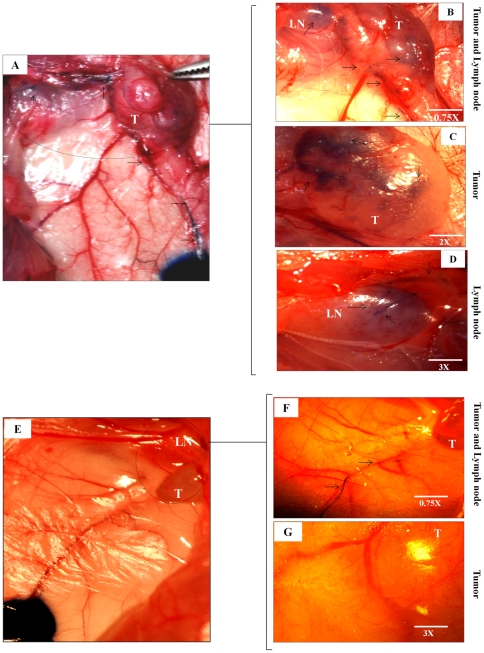
Figure 10. VEGF-D knock down reduced lymphatic ingrowths into the tumor:
(A) Digital camera captured picture at 5 minutes show the Evans blue dye injection sites and the dye stained lymphatics going towards the 468LN tumor (T). (B) Images were captured at 15 minutes using a dissection microscope. These images show lymphatics (arrows) going into the 468LN tumors as well as tumor draining axillary lymph nodes (LN). (C) Image of same tumor at a higher magnification (see inset) captured at 20 minutes showing blue dye inside the tumor (T), and (D) inside the lymph node (LN) at a higher magnification (see inset). (E) In contrast, VEGF-D knock down (ΔVEGF-D/468LN) tumor implants showed very few lymphatics traceable from the injection site captured with a digital camera. (F) Picture taken at 15 minutes showing lymphatics alone but very little dye in the tumor. (G) Even at 25 minutes no dye-stained lymphatics was visible on the surface or inside the tumor at a higher magnification (see inset).

