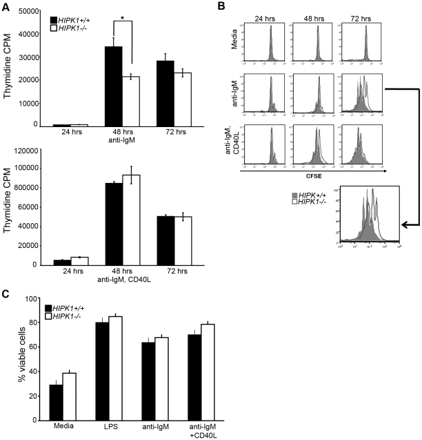
Figure 8. HIPK1 is required for BCR-induced proliferation.
A, Proliferation of HIPK1+/+ and HIPK1−/− splenic B cells in response to anti-IgM ± CD40L (10 µg/ml each) was assessed by [3H]-thymidine incorporation. Cultures were pulsed with 1 µCi tritiated thymidine 12 hrs before the indicated time points. Representative results of three independent experiments are shown. B, Cell division of HIPK1+/+ and HIPK1−/− splenic B cells was determined by CFSE dilution assay. Cells were stimulated with anti-IgM ± CD40L (10 ug/ml) or media alone, and analyzed at 24, 48, and 72 hrs post-stimulation. The solid peaks are wild type and the empty peaks are HIPK1−/−. FACS plots are representative of three independent experiments. C, Viability was measured by FACS by gating on the AnnexinV and PI double-negative populations at 48 hrs after stimulation (n = 4). *p≤0.05.

