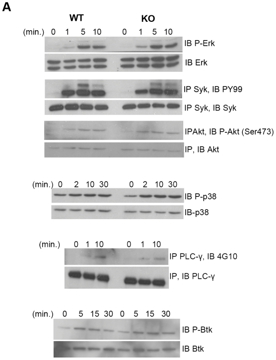
Figure 10. BCR signaling in the absence of HIPK1.
A, Primary B cells were stimulated with anti-IgM (10 µg/ml) for the indicated amounts of time and then lysed. Lysates were subjected to SDS-PAGE and were probed with phospho-specific antibodies as indicated. Blots are representative of minimally three independent experiments.

