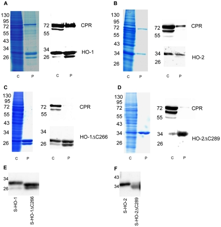
Figure 3. Coomassie staining and immunoblot analysis of co-infections of HO-isoforms and CPR in Sf9 cells.
(A) S-HO-1/CPR coinfection (B) S-HO-2/CPR coinfection (C) S-HO-1ΔC266/CPR coinfection (D) S-HO-2ΔC289/CPR coinfection. The respective lanes C show cytosolic fractions (80 µg protein) of the co-infections and lanes P samples after purification (5 µg). For purification Strep-tagged HO isoforms were used in combination with untagged CPR. Coomassie staining was used to control the degree of purification. Western blot analysis was performed using specific antibodies against CPR (upper blot) and the HO-isoforms 1 and 2 (lower blot). Panel E and F show a HO-Western-blot analysis for direct comparison of full-length and truncated isoforms. The molecular weight of the proteins was specified in kDa.

