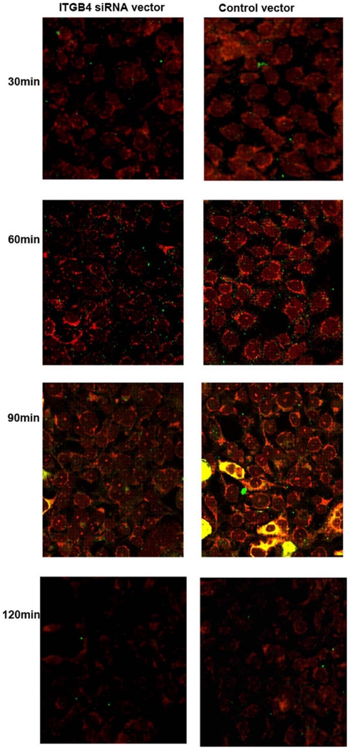
Figure 2. Colocalization of HLA-DR with endocyosed OVA in respiratory epithelial cells.
16HBE14o- cells from different groups were pulsed with FITC-labeled OVA for 0–120 min, fixed, permeabilized, and stained with Dylight 549-labeled anti-HLA-DR antibodies. Cells were analyzed by confocal microscopy. Yellow staining (red+green) indicates colocalization. Results are representative of 1 experiment repeated 3 times.

