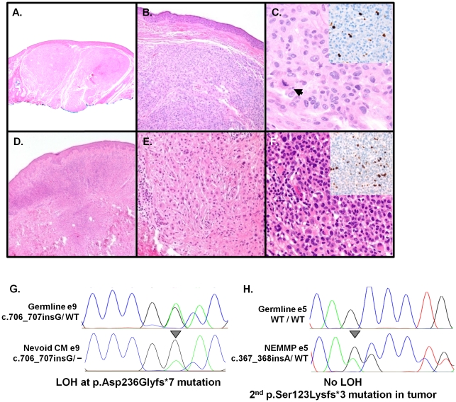
Figure 2. Histologic and molecular analyses of tumors from Fam-562.
(A)–(F): Histology of 2 distinct NEMMPs. (A) Scanning view of the first lesion showing two expansile dermal nodules (H&E, 2×,) with a (B) benign nevoid appearance (H&E, 10×). (C) Atypical cytological features including nuclear pleomorphism and prominent nucleoli and a dermal mitotic figure (arrow) (H&E, 40×) along with focal increases in Ki67 staining (inset). (D) In the second lesion, there is an expansile dermal proliferation (H&E, 4×). (E) Detail of a field populated by dermal nevic cells with bland nuclear features (H&E,20×). (F) A proliferative area showing marked nuclear atypia and hyperchromasia along with elevated Ki 67 staining (inset). Biallelic inactivation of BAP1 in two tumors through (G) loss of the wildtype allele in a nevoid melanoma (ie. LOH; arrow) or (H) a secondary mutation (p.Ser123Lysfs*3) in a NEMMP that did not exhibit LOH.

