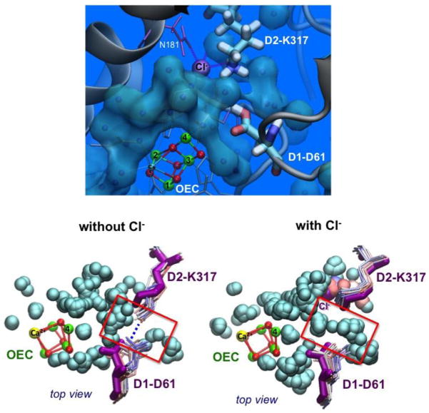
Fig 3.
Top: Waters modeled in the 1.9 Å X-ray structure (gray spheres) next to the Cl−, OEC, and residues D1-Asp61 (D61) and D2-Lys317 (K317). Bottom: Superposition of instantaneous configurations along MD simulations (waters shown as gray spheres and D61, K317 side chains colored from red to blue for 0–24 ns) of the OEC with (right) or without (left) Cl− at the BS2 site. A salt-bridge between K317 and D61 forms upon Cl− depletion, and is interrupted by water in the presence of Cl−. The X-ray configuration is shown in magenta. Reproduced from Ref. 29**.

