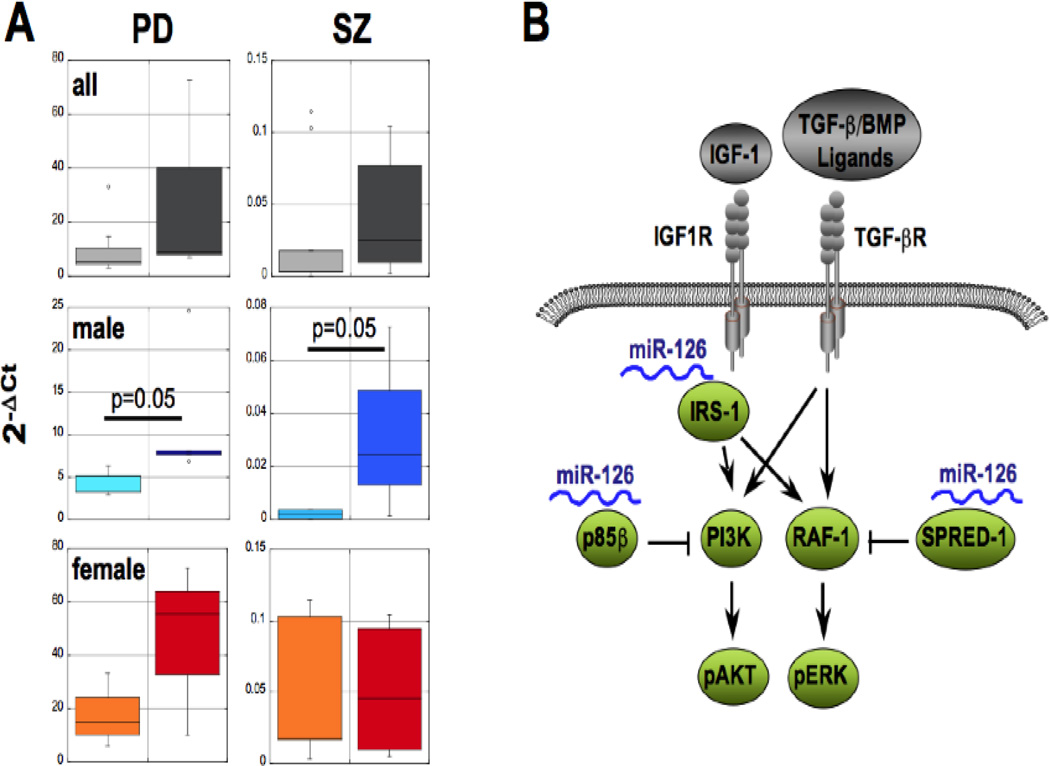
Figure 1.
Expression of miR-126 in PD and schizophrenia (SZ) and its targeting of PI3K signaling pathways. (A) Substantia nigra DA neurons from sporadic PD patients (5 males and 3 females, right bars) or pyramidal neurons in layer 3 of the superior temporal gyrus (STG) of the cerebral cortex from SZ patients (4 males and 5 females, right bars) and aged-matched controls (PD: 5 males and 3 females, SZ: 4 males and 5 females, left bars) were isolated by laser-assisted microdissection from postmortem brain material as previously described (Pietersen, et al., 2011). miRNA expression profiles were performed by Megaplex Human miRNA TaqMan® Arrays (LifeTechnologies) and quantified by the 2−ΔCt method (Livak and Schmittgen, 2001) using RNU44 or MammU6 snoRNA expression for normalization. Data are plotted for all samples combined or males and females separately, and statistical significance determined by 3-way ANOVA and student’s t-test. (B) Schematic of upstream factors in IGF-1/TGF-1β/PI3K signaling and validated targets of miR-126.

