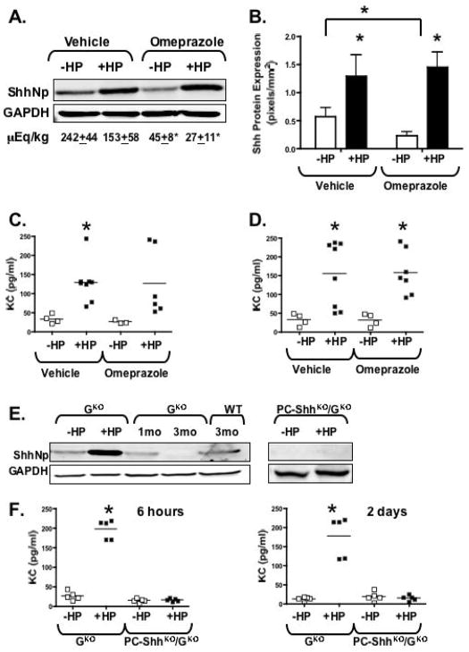
Fig. 3. Shh protein expression in response to acute H. pylori infection in hypochlorhydric omeprazole-treated controls and gastrin-deficient mice.
(A) Representative western blot of Shh protein (ShhNp, 19kDa processed Shh) expression in stomach homogenates collected from vehicle- or omeprazole-treated mice without H. pylori (−HP) or with H. pylori (+HP) 6 hours post-infection. Acid levels measured as μEq/kg of H+ are shown. (B) Quantification of Shh protein expression. Data are shown as means ± SEM for 3 individual experiments and expressed as Shh (pixels/mm2), *P < 0.05 compared to uninfected mice. Tissue KC concentrations measured by Luminex® multiplex assay in stomach collected from vehicle- or omeprazole-treated mice without H. pylori (−HP) or with H. pylori (+HP) 6 hours (C) and 2 days (D) post-infection. (E) Representative western blot of ShhNp expression in gastrin-deficient (GKO) and PC-ShhKO/GKO mice without H. pylori (−HP) or with H. pylori (+HP) 6 hours post-infection. Also shown are changes in Shh protein expression in 1 and 3 month old GKO and 3 month old wild type (WT) mice. (F) Tissue KC concentrations measured by Luminex® multiplex assay in stomach collected from gastrin-deficient (GKO) and PC-ShhKO/GKO mice without H. pylori (−HP) or with H. pylori (+HP) 6 hours and 2 days post-infection. n = 4–7 per group, *P < 0.05 compared to uninfected mice analyzed by two-way ANOVA.

