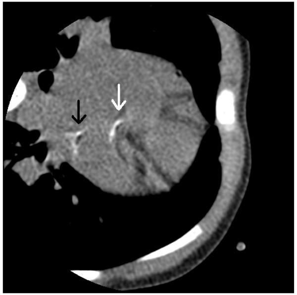
Figure 1.
CT angiogram of the heart prior to contrast. There is abnormal calcification on the left circumflex artery (white arrow) and left main coronary artery (black arrow). There is normal calcification in the sternum and ribs. Photo courtesy of Dr. Sara Arnold at Children’s Hospital of Wisconsin.

