Fig 1.
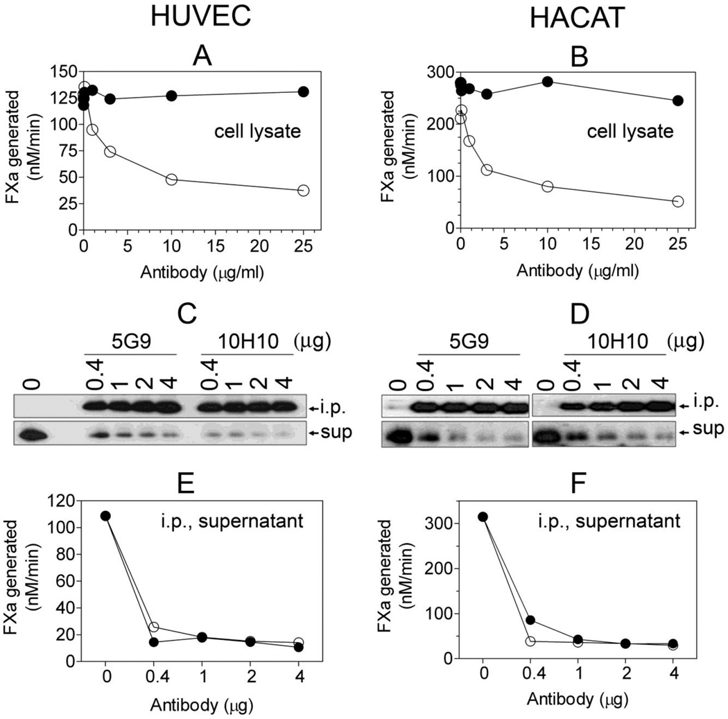
TF mAb 10H10 does not inhibit TF procoagulant activity but efficiently depletes the procoagulant TF from cell lysates following immunoprecipitation. Human umbilical vein endothelial cells (HUVEC) and human keratinocytes (the HaCaT cell line) were cultured as monolayers in 100 mm culture dishes. HUVEC were stimulated with TNFα and IL-1β (20 ng/ml each) for 6 h. The cells were harvested in HEPES buffer containing 50 mM octylglucopyranoside, 1 ml/dish for HUVEC and 4 ml/dish for HaCaT). A and B: cell lysates were incubated with varying concentrations of TF mAb 5G9 or 10H10 for 1 h and the residual TF activity was analyzed in FX activation assay after adding FVIIa (10 nM) and FX (175 nM); C and D: cell lysates (200 µl) were incubated with the indicated amounts of TF mAb 5G9 or 10H10 for 2 h, the immunocomplexes were captured on protein G-agarose beads, and TF in immunoprecipitates (i.p.) or depleted supernatants (sup) was detected by Western blotting using a TF polyclonal antibody; E and F, residual TF activity in i.p. depleted supernatants was measured with FVIIa (10 nM) and FX (175 nM). Cell lysates and immunodepleted supernatants were diluted 10 to 100-fold for measuring TF activity. The symbols are: (○), 5G9; (●), 10H10.

