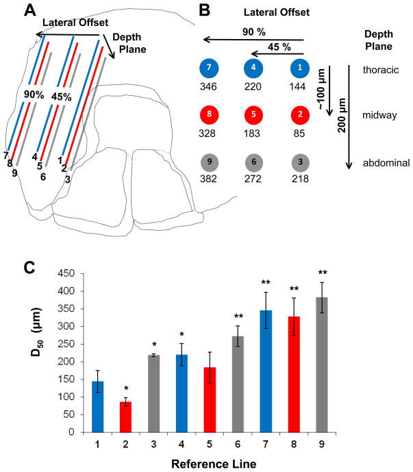
Figure 6. Independent reference lines for nerve position.
A. Nine reference lines were positioned through the thickness (~200 μm) and across the width (~8 mm) of the left costal region of each diaphragm muscle studied in C57BL/6 mice; each line represented 75% of actual nerve length for that muscle region. Through the muscle thickness, reference lines represent three planes: adjacent to thoracic surface (corresponding to the nerve plane), midway through the muscle thickness (corresponding to the arteriolar plane) and adjacent to abdominal muscle surface (deepest plane relative to nerve plane). Within each reference plane, lines were positioned at 45% and 90% of the distance (Lateral Offset) to the costal muscle boundary. B. End view illustrates position of reference lines within muscle boundaries with mean respective D50 values determined according to Figure 5. For actual nerves, the D50 value was 155 ± 30 μm. C. Histogram showing D50 values calculated for each reference line. *P<0.05 vs. actual nerves; ** P<0.01 vs. actual nerves.

