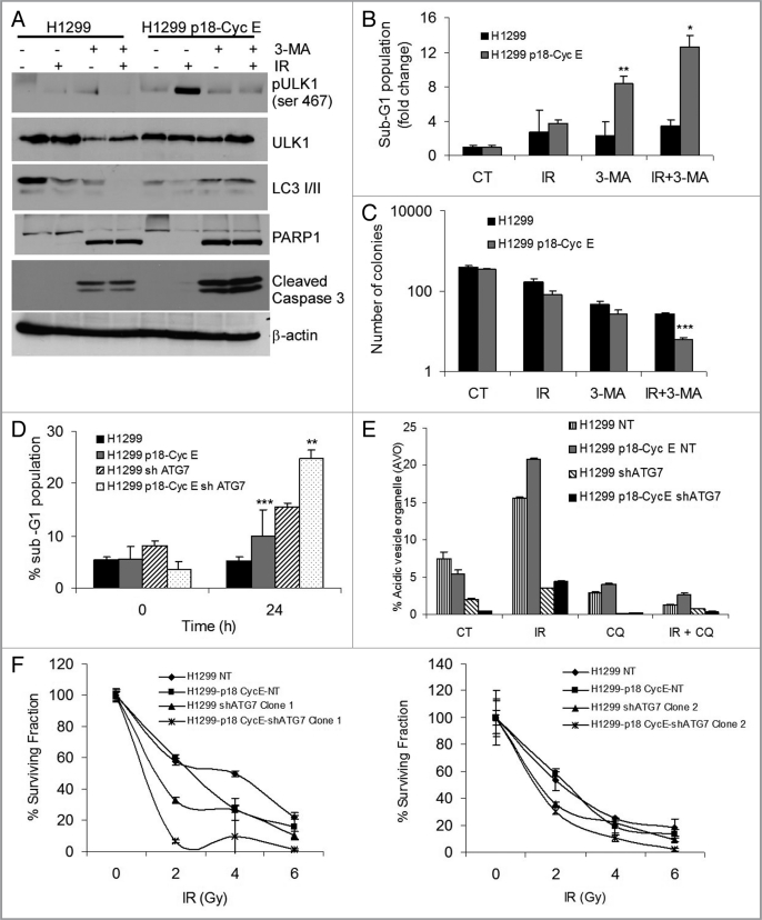
Figure 5.
Autophagy inhibition increases apoptosis in p18-CycE-expressing cells. (A) Cells harvested and lysed at 24 h post IR in the absence or presence of 3-MA (10 mM) were immunoblotted for pULK1-ser467, ULK1, LC3 I/II, PARP1, cleaved caspase-3, and β-actin. (B) Cell death at 24 h following IR in the absence or presence of 3-MA (10 mM) treatment is shown as percentage of cells with sub-G1 DNA content. **p = 0.02, *p = 0.03 Student's t-test. (C) Clonogenic assay following IR (5 Gy) in the absence or presence of 3-MA. *p < 0.001 Student's t-test. (D) Cell death is shown as percentage of cells with sub-G1 DNA content that stably express p18-CycE in cells without or with shATG7 expression (Clone 1) at 24 h following IR. *p < 0.001, **p = 0.001 Student's t-test. (E) Detection of acidic vesicular organelles (AVOs) by measuring red and green fluorescence of acridine orange-stained cells using FACS analysis in parental and p18-CycE expressing cells with or without shATG7 expression following IR and/or chloroquine (100 µM) treatment for 24 h. Data represent means ± s.e.m., obtained from three independent triplicates. (F) Clonogenic assay for cells stably-expressing p18-CycE in the absence or presence of shATG7 (Clone 1 in left panel, Clone 2 in right panel) following IR. p < 0.05 Student's t-test. For all panels, values are mean ± s.e.m. of three independent experiments performed in triplicates.

