Figure 2.
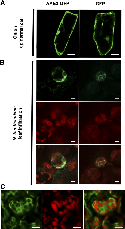
Subcellular Localization of AAE3.
(A) GFP fluorescence images of onion epidermal cells transiently expressing AAE3-GFP (left panel) and free GFP (right panel). Bars = 50 μM.
(B) Transient expression of an AAE3-GFP fusion (left panels) and free GFP (right panels) in N. benthamiana leaf parenchyma cells introduced by Agrobacterium infiltration. GFP fluorescence images (top panels), chloroplast autofluorescence (middle panels), and merge of GFP and autofluorescence (bottom panels). Bars = 5 μM.
(C) Confocal image displaying the upper focal plane of mesophyll cells in Arabidopsis plants expressing a AAE3p:AAE3-GFP transgene. GFP fluorescence image (left panel), chloroplast autofluorescence (middle panel), and merge of GFP and autofluorescence (right panel). Bars = 10 μM.

