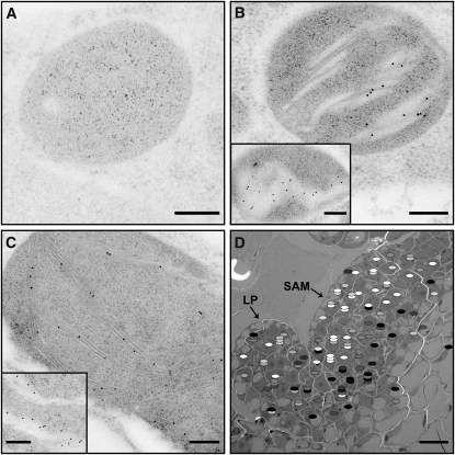
Figure 7.
Immunogold Labeling of OEC33 in the Shoot Apex.
(A) to (C) Images of plastids from the CZ of the L2 layer (A), the L3 periphery (B), and of a chloroplast from a relatively mature leaf primordium (C), labeled with an antibody against subunit 33 of the OEC. No labels are present in the plastid from the center of L2 ([A]; the small dark spots seen in the image are stain particles), consistent with the absence of thylakoid membranes in this region. By contrast, multiple labels, localized to the thylakoid membranes, are seen in the plastids from the L3 layer (B) and the primordial leaf (C). Labeling is more pronounced in sections made along the thylakoid sheet plane (insets in [B] and [C]), which expose the luminal compartment, where the OEC resides. Bars = 200 nm.
(D) Map summarizing the label density of plastids of the SAM and a young leaf primordium. Each ellipse represents one plastid and stacked ellipses represent plastids belonging to the same cell. The density of label is represented in grayscale (from white [low] to black [high]) and normalized to that of older leaf chloroplasts present in the same section. As no averaging was made, and because the number of plastids and labels was relatively small, the data are noisy. Nevertheless, the pattern emerging is consistent with the structural and chlorophyll fluorescence data. LP, leaf primordia. Bar = 10 μm.

